

johndoe@gmail.com
Communication Disorders
By Louise Cummings
The website is a vitally important resource for student readers and for practitioners wishing to develop their knowledge. It contains 200 multiple choice questions and answers which examine every disorder discussed in the book. There is also an extensive array of audio and visual material on the website that covers everything from the speech of people who clutter to the numerous organic pathologies that cause voice disorders.
Throughout the text you will find boxes indicating that there is complementary material available on the website. All material is organized by chapter to make it easy to navigate. We hope you find the materials on this website interesting and useful.
Click on the following links to go straight to their related resources:
MCQs with answers | Chapter 1 | Word Document | (0.48Mb)
MCQs with answers | Chapter 2 | Word Document | (0.48Mb)
MCQs with answers | Chapter 3 | Word Document | (0.48Mb)
Audio file of two speakers (a man and a woman) who have undergone glossectomy
Chapter 4 | Audio (mp3) | (2.13Mb)
MCQs with answers | Chapter 4 | Word Document | (0.48Mb)
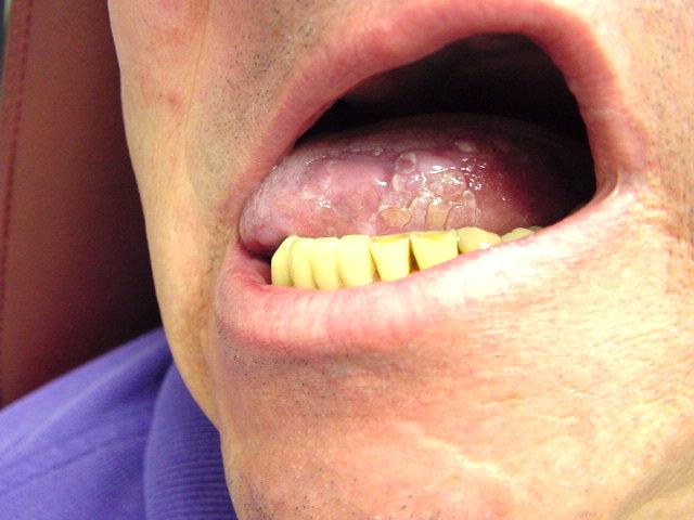
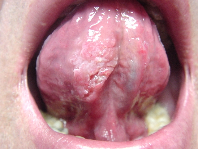
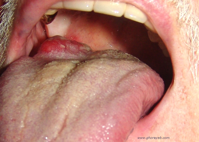
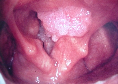
Images of carcinoma of the tongue
Four images of carcinoma of the tongue (Reproduced with kind permission of Bechara Y. Ghorayeb, MD; www.houstonoto.com/PicturesLarynx.html)

MCQs with answers | Chapter 5 | Word Document | (0.48Mb)
MCQs with answers | Chapter 6 | Word Document | (0.49Mb)
Selective mutism (reproduced with kind permission of RDF Television and Channel 4)
Pre- and post-surgical voices of speakers who have undergone cricothyroid approximation (reproduced with kind permission of James P. Thomas, MD, www.voicedoctor.net).Audio
These audio files can be accessed from:
http://voicedoctor.net/surgery/audio-examples?f[0]=field_type_audio:141
Audio file of a baby boy with cri-du-chat syndrome (reproduced with kind permission of the Cri Du Chat Support Group of Australia)
Chapter 7 | Audio (mp3) | (0.7Mb)
Audio file of a baby girl with cri-du-chat syndrome (reproduced with kind permission of the Cri Du Chat Support Group of Australia)
Chapter 7 | Audio (mp3) | (1.81Mb)
Audio file relating to Exercise 7.3 - This audio file is reproduced with kind permission of The National Association of Laryngectomee Clubs (www.laryngectomy.org.uk)
Audio (mp3) | (2.83Mb)
MCQs with answers
Chapter 7 | Word Document | (0.48Mb)
Multiple Sclerosis - Audio file reproduced with kind permission of Richard Stasney, MD of the Texas Voice Center (www.texasvoicecenter.com).
Audio (mp3) | (1.18Mb)
Myasthenia Gravis - Audio file reproduced with kind permission of Richard Stasney, MD of the Texas Voice Center (www.texasvoicecenter.com).
Audio (mp3) | (1.27Mb)
Parkinson's Disease 1 - Audio file reproduced with kind permission of Richard Stasney, MD of the Texas Voice Center (www.texasvoicecenter.com).
Audio (mp3) | (1.57Mb)
Parkinson's Disease 2 - Audio file reproduced with kind permission of Richard Stasney, MD of the Texas Voice Center (www.texasvoicecenter.com).
Audio (mp3) | (1.18Mb)
Puberphonia 1 - Audio file reproduced with kind permission of Richard Stasney, MD of the Texas Voice Center (www.texasvoicecenter.com).
Audio (mp3) | (0.79Mb)
Puberphonia 1 post voice tx - Audio file reproduced with kind permission of Richard Stasney, MD of the Texas Voice Center (www.texasvoicecenter.com).
Audio (mp3) | (0.79Mb)
Puberphonia 2 - Audio file reproduced with kind permission of Richard Stasney, MD of the Texas Voice Center (www.texasvoicecenter.com).
Audio (mp3) | (1.58Mb)
Puberphonia 2 post voice tx - Audio file reproduced with kind permission of Richard Stasney, MD of the Texas Voice Center (www.texasvoicecenter.com).
Audio (mp3) | (1.41Mb)
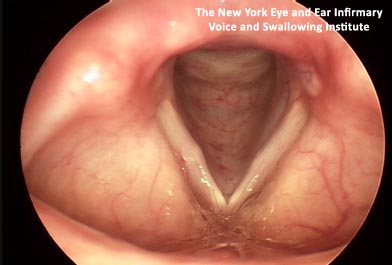
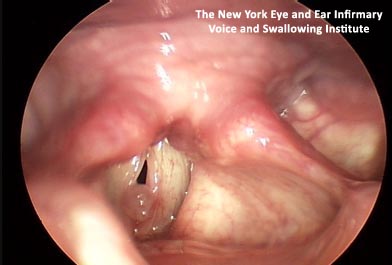
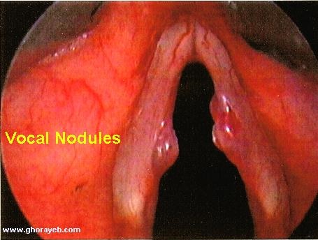
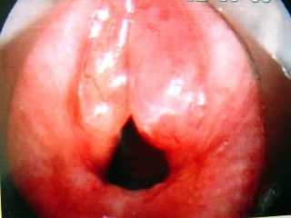
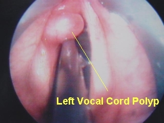
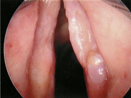
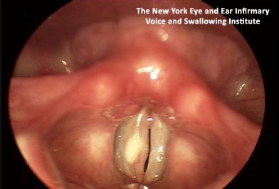
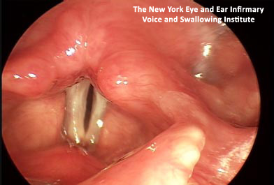
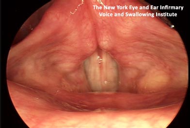
Images of vocal nodules and polyps
Images reproduced with kind permission of The New York Eye and Ear Infirmary Voice and Swallowing Institute (www.nyee.edu/vsi):
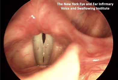
Nodules The nodules prevent the vocal folds from closing completely during phonation.
The remaining images in this section are reproduced with kind permission of Bechara Y. Ghorayeb, MD (www.houstonoto.com/PicturesLarynx.html):
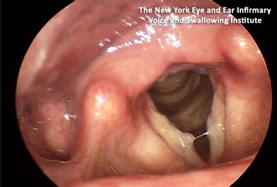
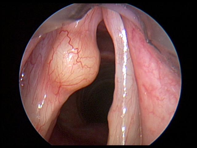
Images of vocal cysts
These images are reproduced with kind permission of The New York Eye and Ear Infirmary Voice and Swallowing Institute (www.nyee.edu/vsi):

This image is reproduced with kind permission of Bechara Y. Ghorayeb, MD (www.houstonoto.com/PicturesLarynx.html).
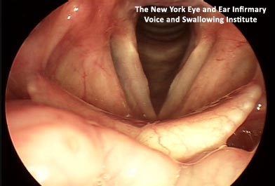
Images of Reinke’s oedema and granuloma
These images are reproduced with kind permission of The New York Eye and Ear Infirmary Voice and Swallowing Institute (www.nyee.edu/vsi):
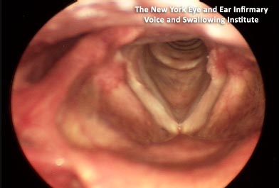
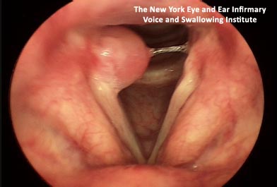
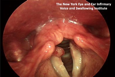
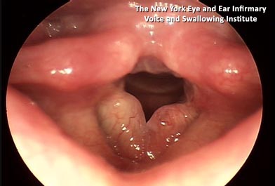
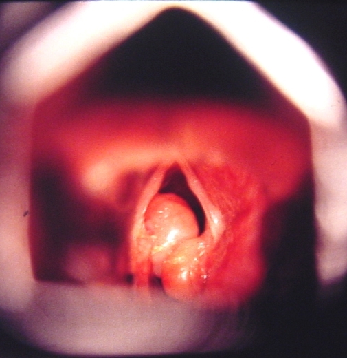
Image reproduced with kind permission of Bechara Y. Ghorayeb, MD
(www.houstonoto.com/PicturesLarynx.html).
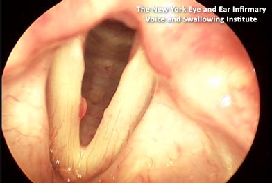
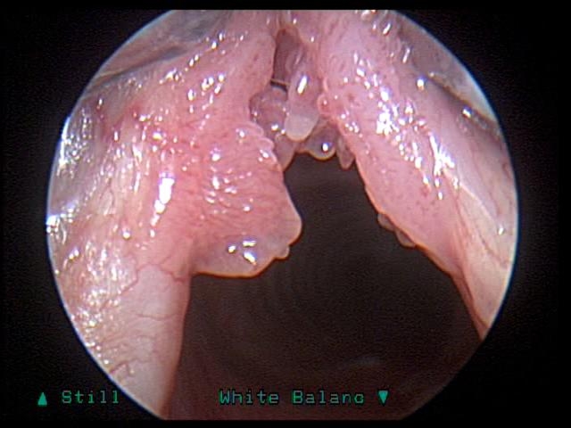
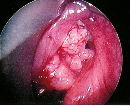
Images of laryngeal papillomas
Images reproduced with kind permission of Bechara Y. Ghorayeb, MD
(www.houstonoto.com/PicturesLarynx.html):
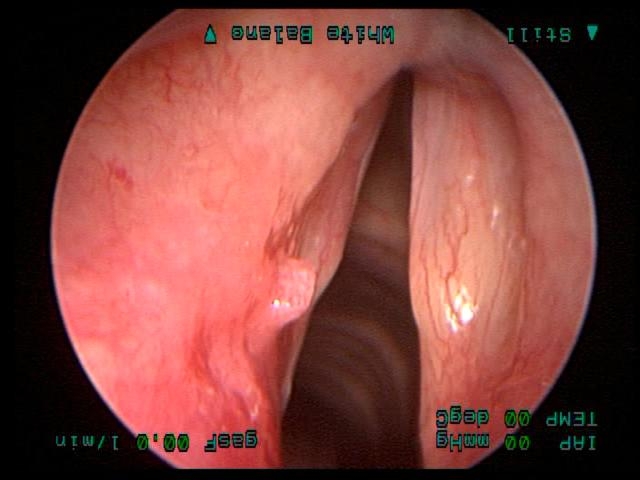
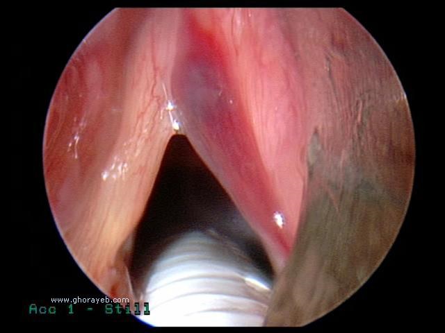
Sulcus The sulcus on each vocal fold can be seen as a ridge running the length of the folds.
Audio file of presbylarynx
This audio file is linked with kind permission of Richard Stasney, MD of the Texas Voice Center (www.texasvoicecenter.com). It can be accessed at: www.texasvoicecenter.com/diseases.html (presbylaryngis)
Audio files of vocal fold paralysis and paresis
These audio files are linked with kind permission of Richard Stasney, MD of the Texas Voice Center (www.texasvoicecenter.com). They can be accessed at: www.texasvoicecenter.com/diseases.html (unilateral vocal fold paralysis and bilateral vocal fold paralysis)
Audio files of spasmodic dysphonia
These audio files are linked with kind permission of Richard Stasney, MD of the Texas Voice Center (www.texasvoicecenter.com). They can be accessed at: www.texasvoicecenter.com/diseases.html (adductor spasmodic dysphonia and abductor spasmodic dysphonia)
These audio files are linked with kind permission of the National Spasmodic Dysphonia Association (www.dysphonia.org). They can be accessed at: www.dysphonia.org (adductor spasmodic dysphonia and abductor spasmodic dysphonia)
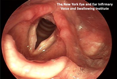
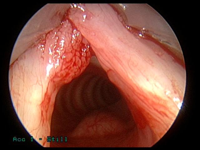
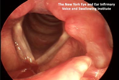
Image of laryngopharyngeal reflux
This image is reproduced with kind permission of The New York Eye and Ear Infirmary Voice and Swallowing Institute (www.nyee.edu/vsi):
Laryngopharyngeal reflux has caused inflammation (redness and swelling) of the back of the larynx. The vocal fold mucosa is irritated and copious thick secretions cover the vocal folds. This causes discomfort and irregular mucosal wave vibration, leading to voice changes.
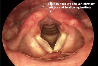
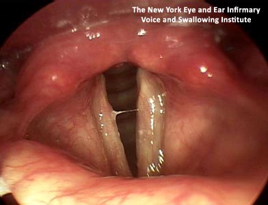
Image of muscle tension dysphonia
This image is reproduced with kind permission of The New York Eye and Ear Infirmary Voice and Swallowing Institute (www.nyee.edu/vsi):
Video file of laryngoscopy examination
This video file is linked with kind permission of James P. Thomas, MD (www.voicedoctor.net). It can be accessed at: www.voicedoctor.net/videos/laryngoscopy-art-seeing-voice-vocal-cords (Laryngoscopy: the art of seeing the voice or vocal cords)
Lecture on cricothyroid approximation
This video file is linked with kind permissionof James P. Thomas, MD (www.voicedoctor.net). It can be accessed at: voicedoctor.net/videos/feminization-laryngoplasty-2005-bologna-italy (A 15-minute video lecture on CTA & feminization laryngoplasty)
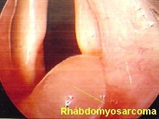
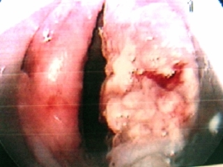
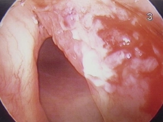
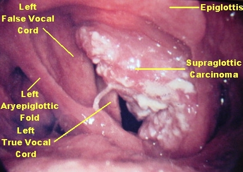
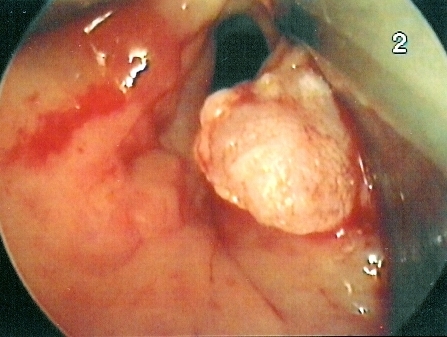
Images of laryngeal cancer
The images below are reproduced with kind permission of Bechara Y. Ghorayeb, MD (www.houstonoto.com/PicturesLarynx.html):
Laryngeal Carcinoma Squamous cell carcinoma of the larynx
Laryngeal Carcinoma Squamous Cell carcinoma
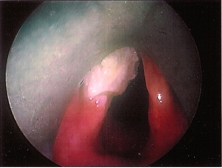
Laryngeal Carcinoma Picture of glottic squamous cell carcinoma of the larynx. The tumor involves the anterior half of the left vocal cord
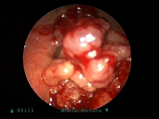
Laryngeal Carcinoma Picture of an extensive squamous cell carcinoma of the larynx. The tumor involves the subglottic region, the glottis and the supraglottic area. This patient underwent a tracheotomy prior to laryngoscopy.
Video files of laryngectomy speakers
Laryngectomy 1
Laryngectomy 2
Videos reproduced with kind permission of The National Association of Laryngectomee Clubs (www.laryngectomy.org.uk).
Chapter 8
Audio file of combined stuttering/cluttering Chapter 8 | Audio (mp3) | (2.79Mb)
MCQs with answers | Chapter 8 | Word Document | (0.51Mb)
Stuttering and Your Child: Help for Parents (reproduced with the kind permission of The Stuttering Foundation - www.stutteringhelp.org)
Help me to Speak – Stammering (reproduced with kind permission of RDF Television and Channel 4)
Let Me Finish: A Stuttering Documentary by Alex R. Murphy
Click here to go to the National Stammering Association website.
Video file of cluttered speech
Video of cluttered speech hosted on the website of the International Cluttering Association http://associations.missouristate.edu/ICA/ To view the video click on ‘What is Cluttering?’ in theassociations.missouristate.edu/ICA/ website.
